Clinical and animal cardioimaging platform
COMMON SUPPORT PLATFORMS
Equipment
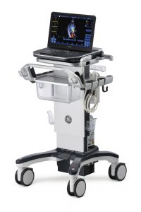
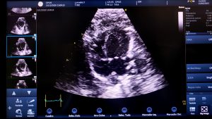
We currently have a high-performance digital ultrasound imaging system Vivid iq with a 12S-RS linear probe (4.5-12.0 MHz) specially configured for cardiac ultrasound in murine models, which allows us to capture images simultaneously with the electrocardiographic recording.
Description
The objective of the unit is to provide professionals in our environment with an effective tool to assess changes in cardiac function in different preclinical models, associated with the pathophysiological process or therapeutic intervention, favoring collaborations and creating synergies between healthcare professionals and researchers.
The ECOCARDIO Clinical and animal cardioimaging platform is made up of a multidisciplinary team with extensive experience in the analysis of cardiac function, from SERGAS, IDIS and USC. Non-invasive imaging techniques are a fundamental piece of current cardiovascular medicine, providing accurate diagnoses and providing prognostic information. In addition, they allow monitoring of different pathophysiological processes, often guiding the therapeutic intervention itself. Echocardiography, due to its wide availability, its low cost, and the absence of radiation, is the most widely used method for the evaluation of cardiac parameters, both at the care and research level.
Services
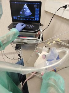
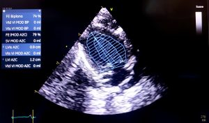
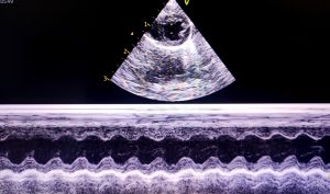
Image capture is performed with the animals lightly anesthetized with isoflurane. Image capture is carried out by performing a parastenal projection of a cut at the level of the papillary muscles using two-dimensional ultrasound following the recommendations of the European and American Societies of Echocardiography, which allows us to evaluate:
• Left ventricular end-diastolic diameter
• Left ventricular end systolic diameter
• Interventricular septal thickness
• Rear wall thickness
• Ejection fraction
• Shortening fraction
Examples:
Fees
VAT not included.
Contact
María Amparo Martínez Monzonís


